Text and Media
Text and Media Headline (max. 100 characters)
- Example
- Example
- Example
- Example
- Example
- Example
- Example
- Example

Text and Media Headline (max. 100 characters)
In this module you can add only text or you can add text and image / video / piechart.
For image and video zoom function is available.
Media items can be left and right aligned.
Also you can add a button.
Pie chart (max. 100 characters)
Here you can see a pie chart.
Multimedia Gallery
Multimedia Gallery (max. 100 characters)
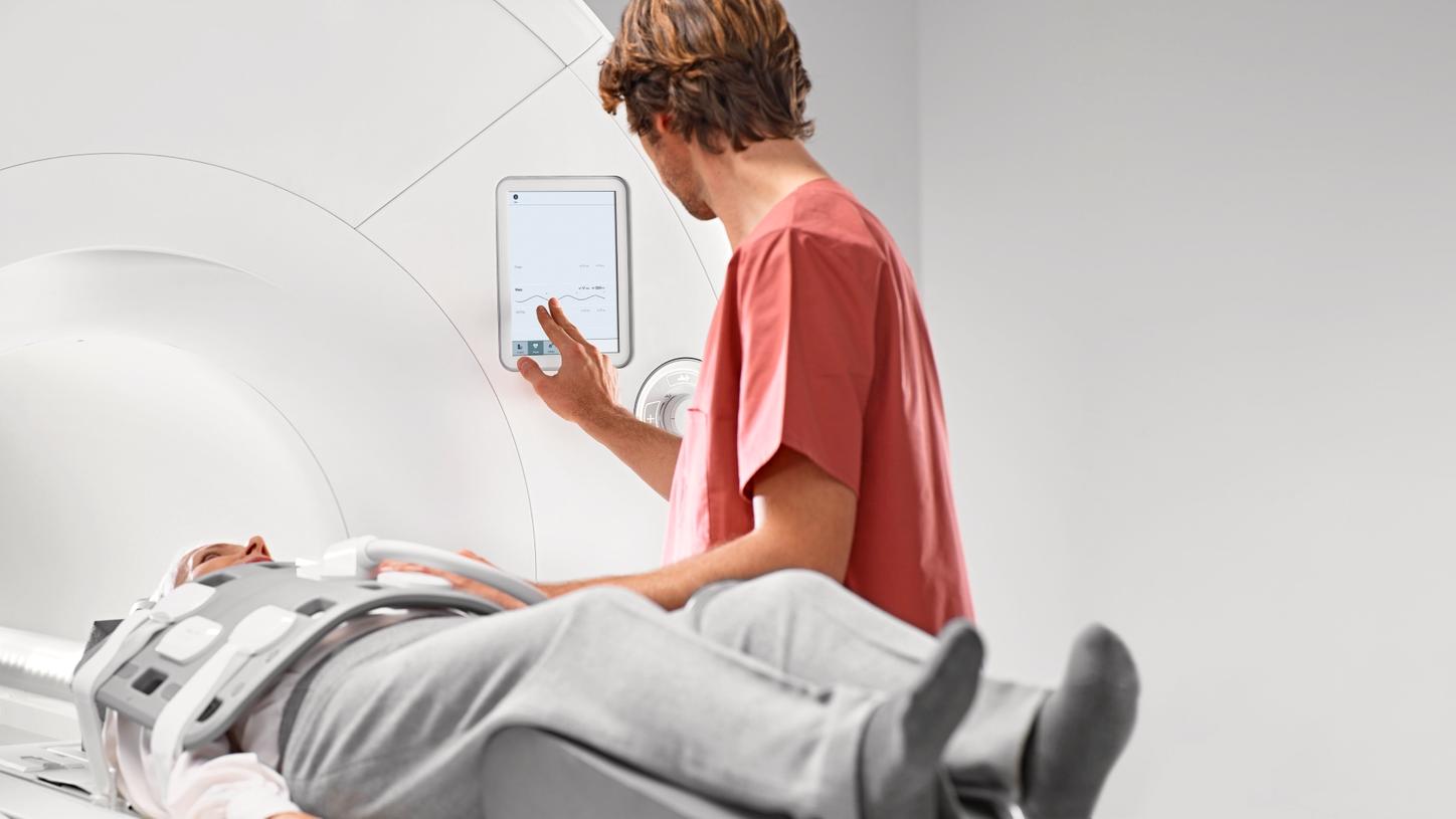
Multimedia Gallery (max. 100 characters)
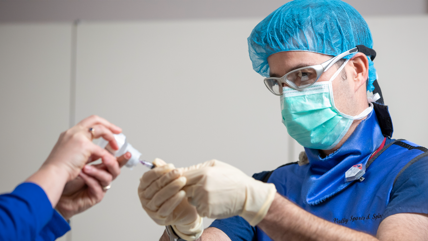
Headline (max. 100 characters)
Headline (max. 100 characters)
Multimedia Gallery (max. 100 characters)
Headline (max. 100 characters)
Headline
Example Clinical image gallery MRI MAGNETOM Free.Max
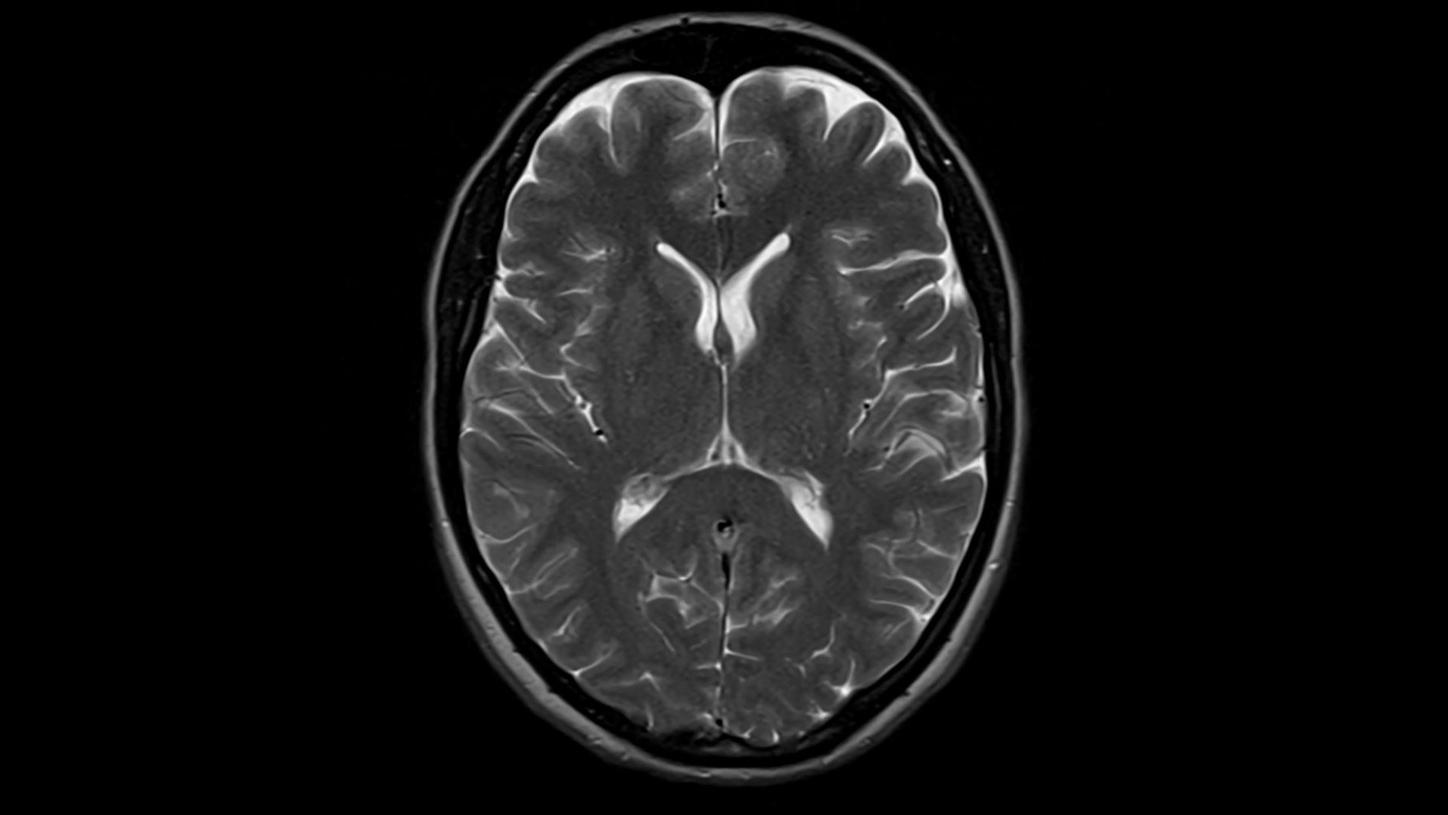
T2 TSE with Deep Resolve
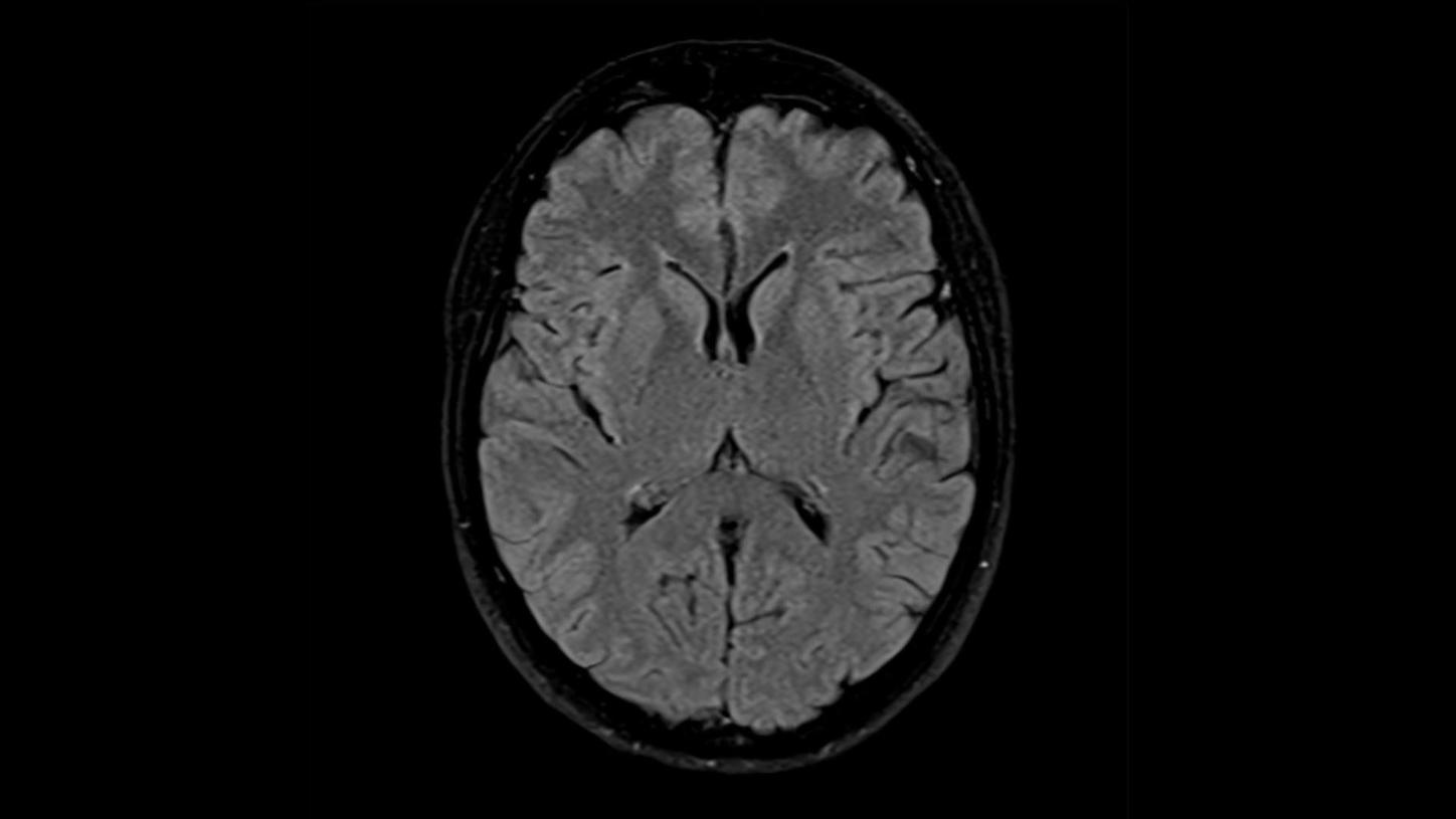
T2 TSE Dark Fluid with Deep Resolve
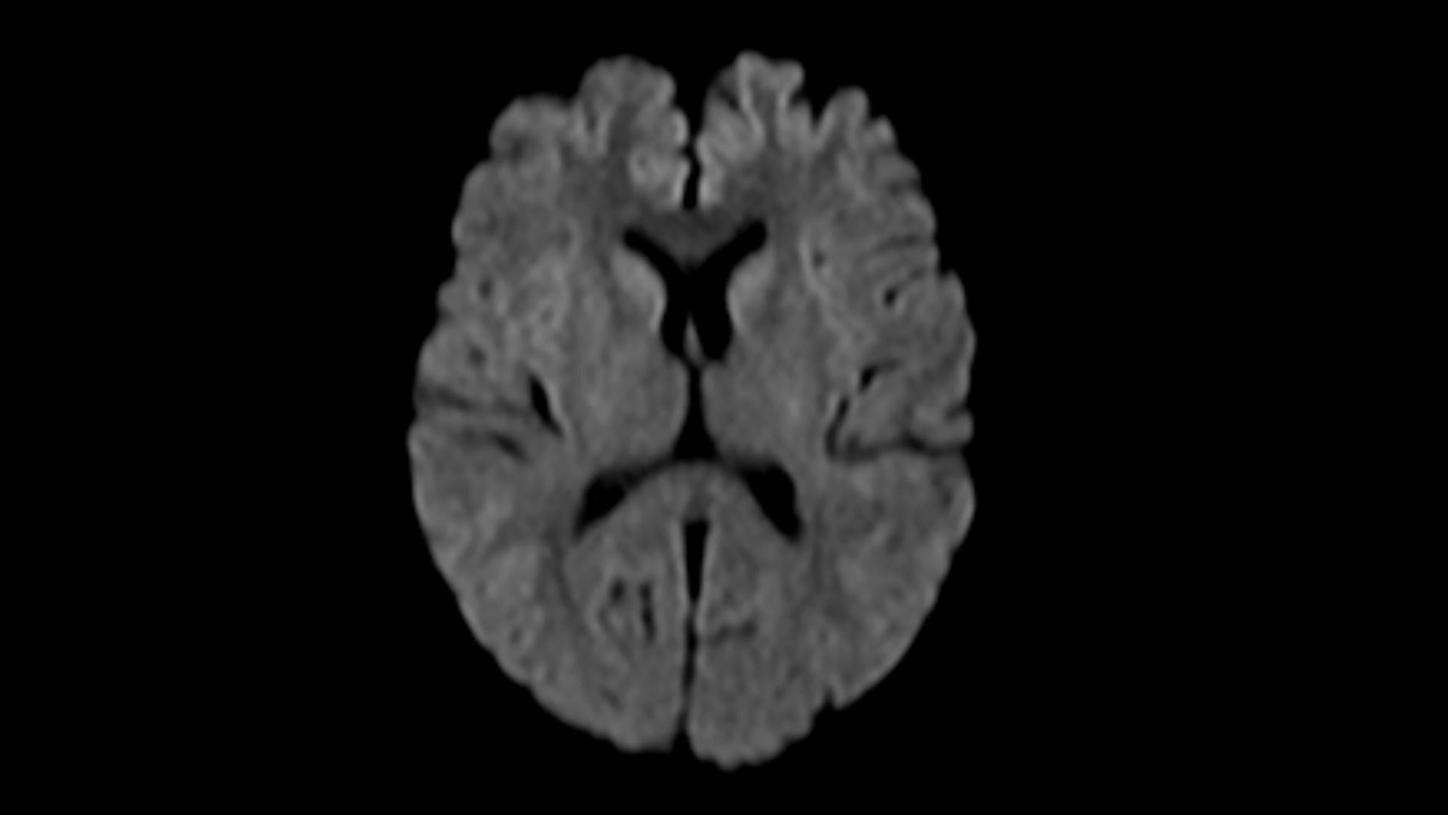
DWI b1000
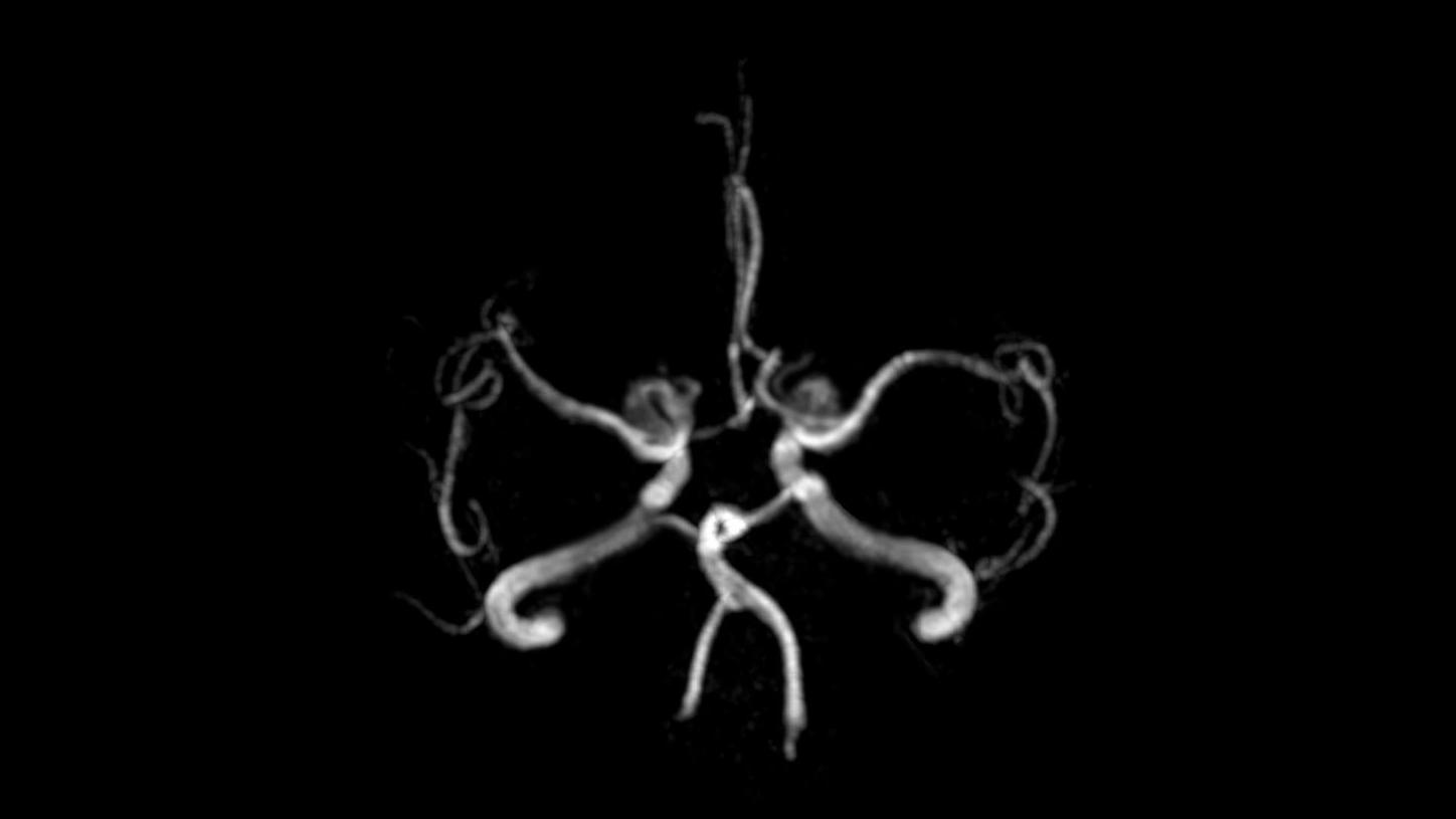
Time-of-flight angiography
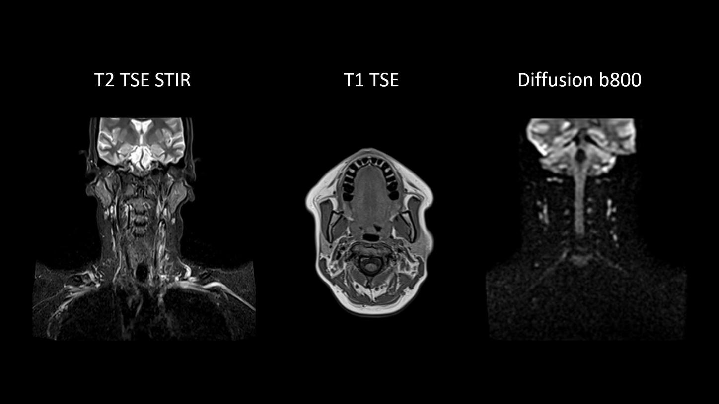
Excellent soft tissue contrast in the neck
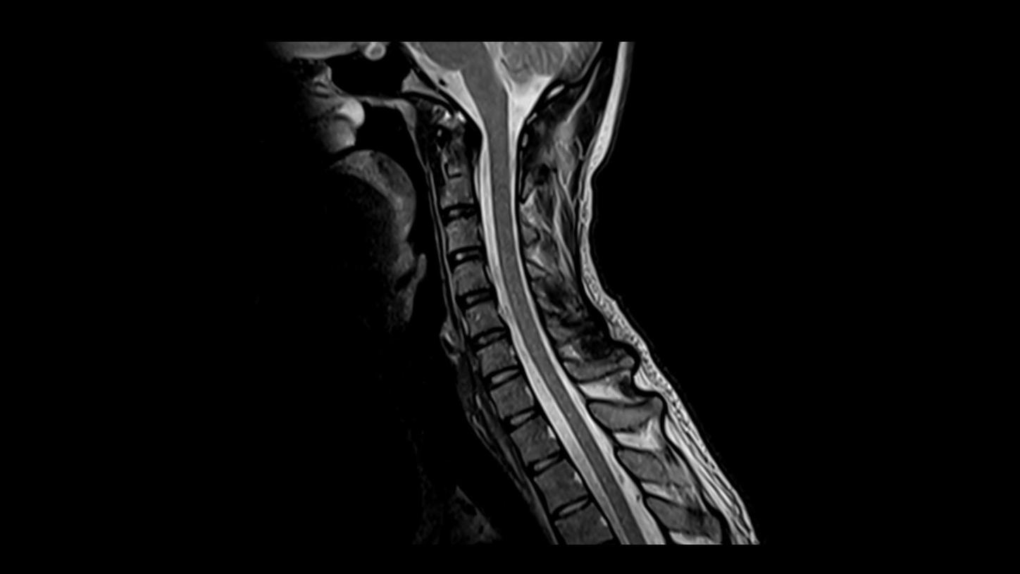
T2 TSE
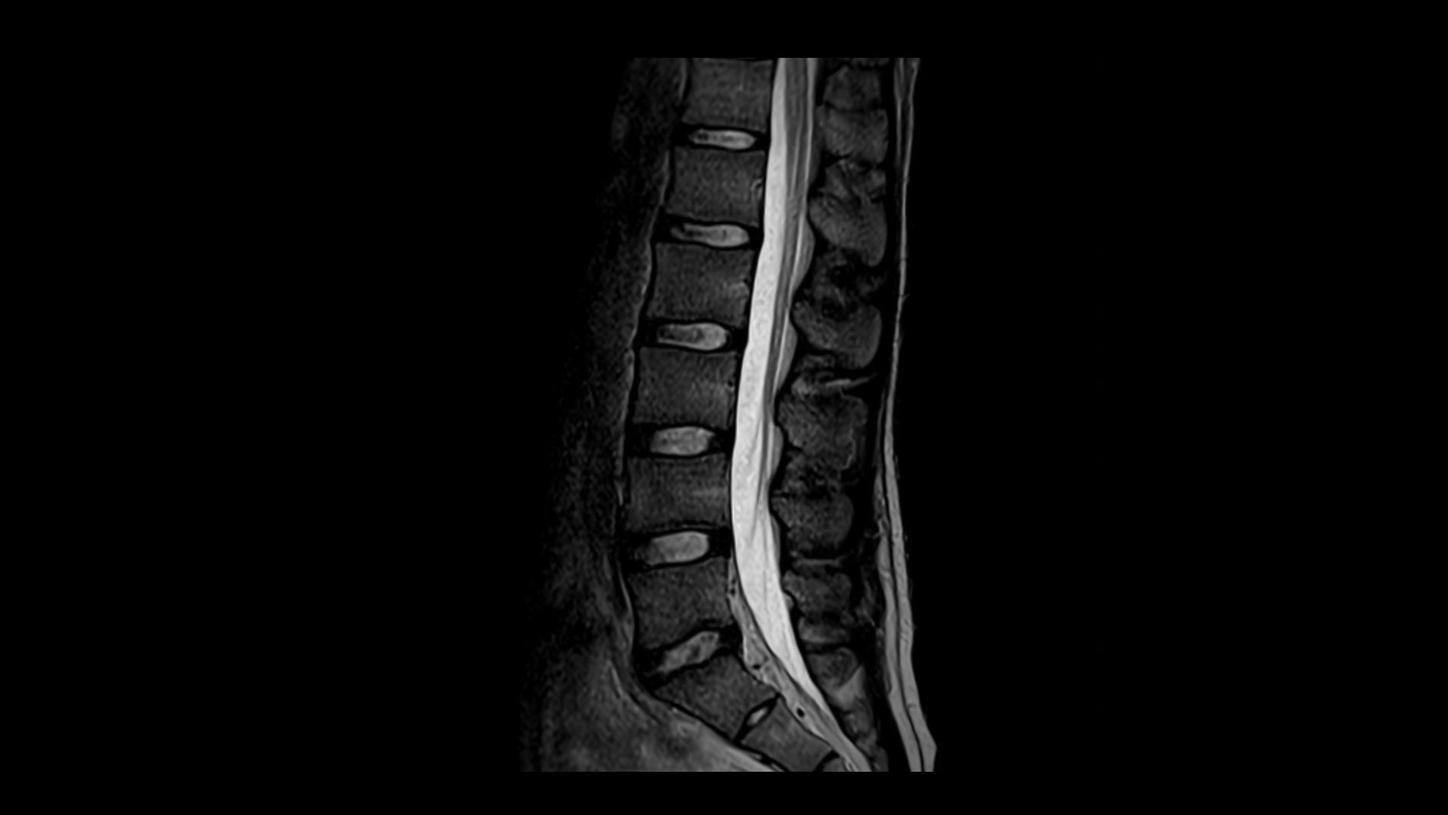
T2 TSE
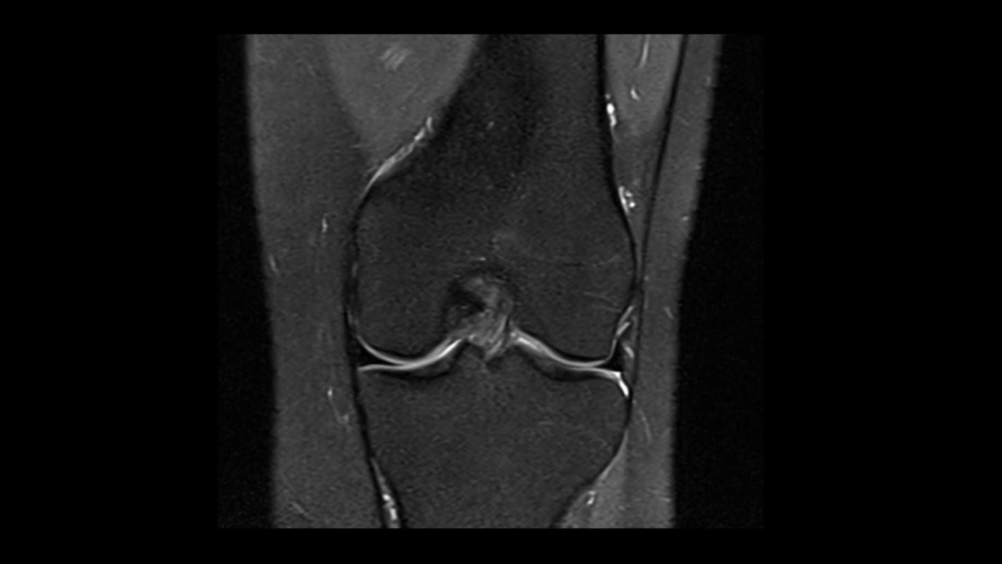
PD TSE Fat Sat with Deep Resolve
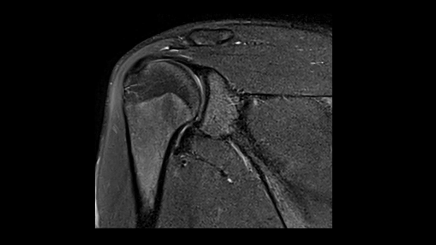
PD TSE Fat Sat with Deep Resolve
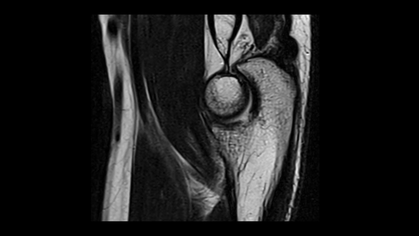
T2 TSE with Deep Resolve

MRCP with Compressed Sensing
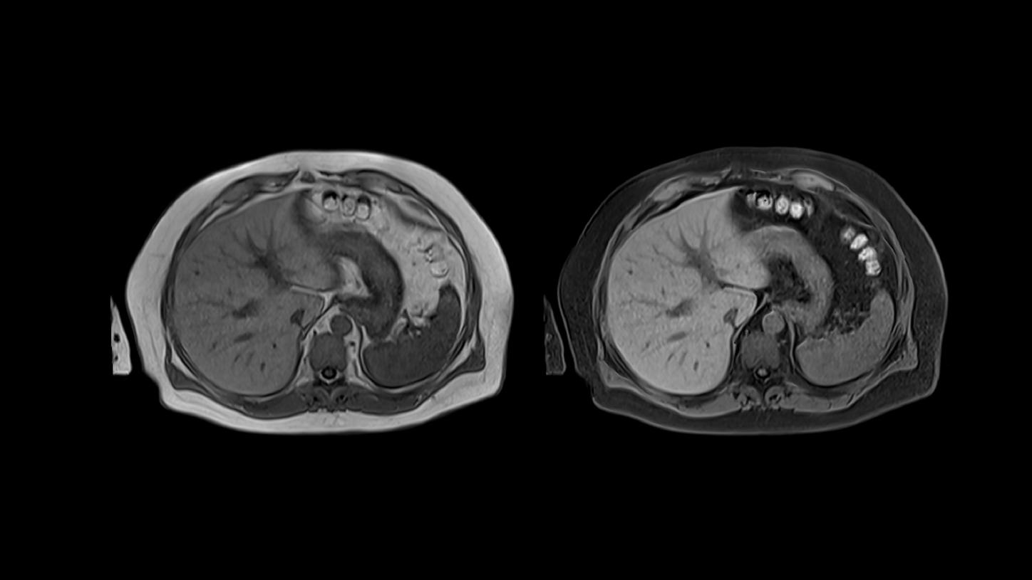
T1 VIBE Dixon with CAIPIRINHA
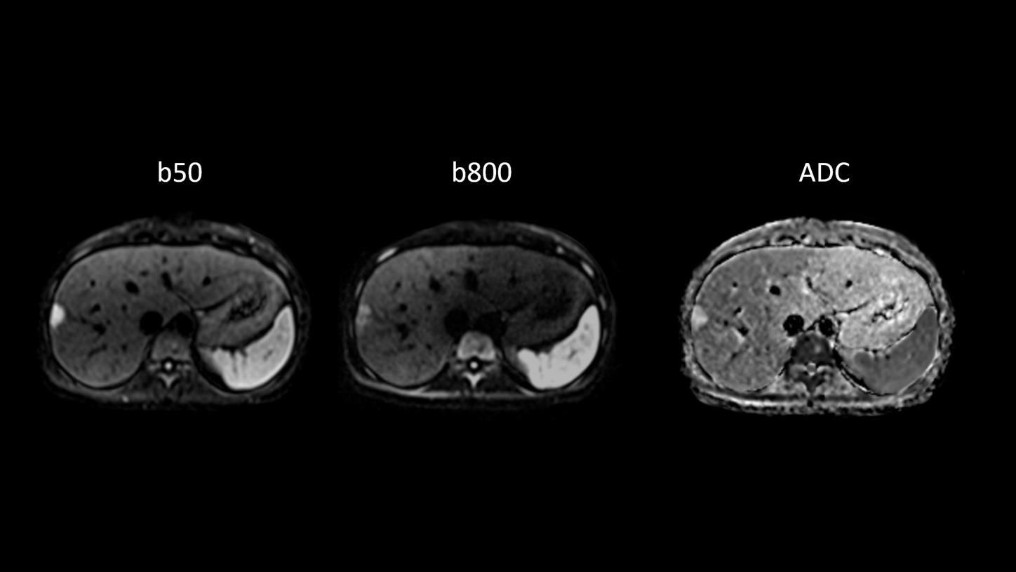
Body diffusion imaging
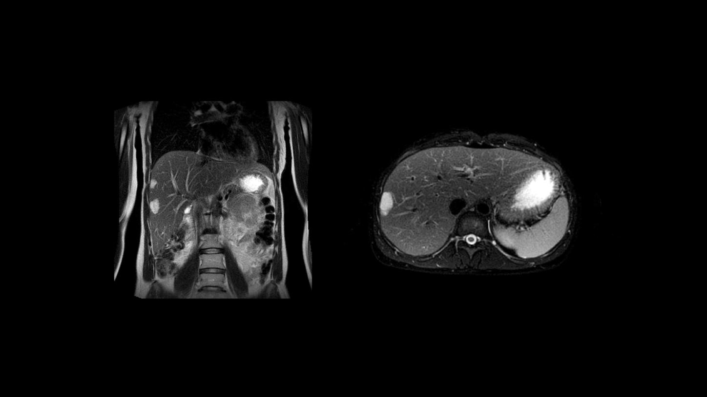
T2 BLADE
Card Column
Card Column: Four cards in a row (max. 100 characters)
You can choose between Text and Media Cards (only image and without zoom function) and Fading Cards (only image and without zoom function). See examples below.
(max. 350 characters)
Text and Media Headline (max. 50 characters)
Description (max. 300 characters)

Text and Media Headline (max. 50 characters)
Description (max. 300 characters)


Card Column: Three cards in a row (max. 100 characters)
(max. 350 characters)
Card Column: Two cards in a row (max. 100 characters)
Description (max. 350 characters)
Text and Media Headline (max. 50 characters)
Description (max. 300 characters)
Text and Media Headline (max. 50 characters)
Description (max. 300 characters)
Quotation Collection
NEW! Quotation Collection (max. 100 chracters)
One to ten quotations possible. It's possible to add an image of the author (image ratio 1x1) as well as an image or logo of the organization. Background is always grey.

This has the potential to redefine clinical decision making right from the first scan. (max. 250 characters)

A completely new way to make small structures of the heart visible.
This will open up the potential to scan more patients on our CT.
In the future every CT will be a photon-counting CT.